2.2. Spatial structure of the
doublet genetic code
For the first time the spatial structure of the
doublet genetic code was proposed in [14].
It is impossible to exclude, that authors were
inspired for construction of this structure by our rhombic variant of the
genetic dictionary [12]
quoted by authors and, in a modified form, even reproduced by them. An alternative doublet code structure was
described also in [3]. The disadvantage of these variants was absence of visual
representation of the symmetry of the two groups of doublets connected
by Rumer’s transformation [13]. Model of spatial structure
of doublet code proposed in our paper [4] eliminates
this defect.
2.2.1. Principles
of model construction
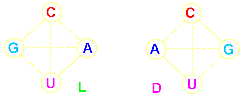
It is possible to construct two types of
connectivity graphs in the form of tetrahedrons of the four nitrogenous bases, in which
a sequence of C, G, U, A, is anti-clockwise (L tetrahedron) and clockwise (D-tetrahedron):
|
|
||
|
It
is possible to allocate three types of single transitions between the bases: |
||
|
C<-->G, U<-->A between the complementary
bases |
C<-->A, U<-->G between non-complementary
bases |
C<-->U, G<-->A transitions of pyrimidine - pyrimidine
and purine
– purine (frequent dotted line) |
|
"Rhombus"
doublets also can be connected with each other by single transitions,
and each neighboring doublet
differs from an initial doublet on one basis, for example CC <--> GC: |
||
|
At the left there are doublets with substitutions in the
first position |
|
On the right there are doublets with substitutions in the second position |
|
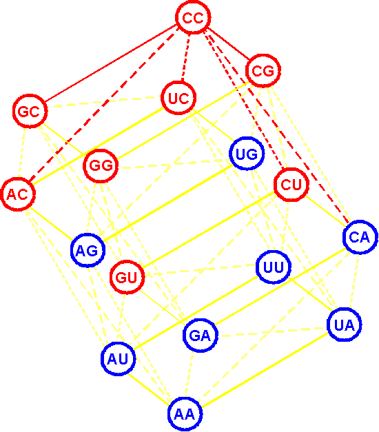
Continuing and further deriving of doublets on
the basis of single substitutions (single
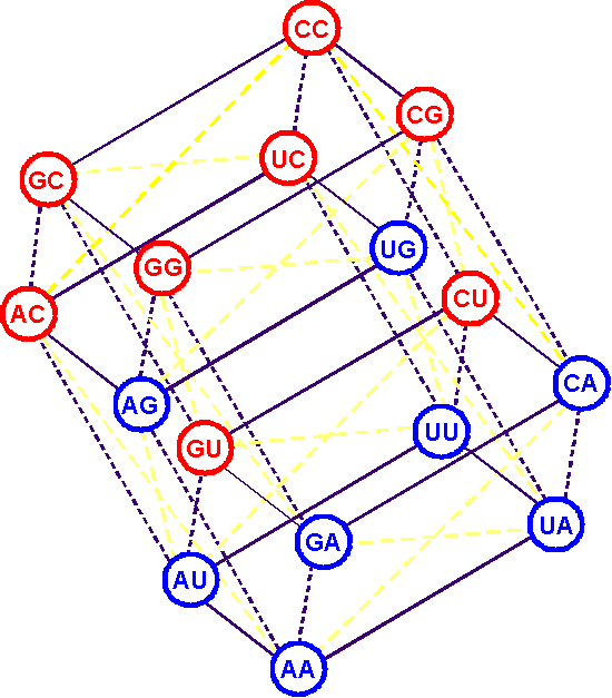
transitions), we finally build the following structure (Fig. 3): |
||
|
We
call it the L-structure. There is also a symmetric D-structure. The resulting L-structure, as mathematicians say, is isomorphic to the
four-dimensional Boolean hypercube (designation B4) superimposed with additional lines along the diagonals of the parallelograms. Such structure is called a "six-dimensional
simplex." Each doublet, located at the
vertex of the hypercube is connected by single transitions with six
neighboring: |
|
Example: 6 lines proceed from doublet СС
connecting 6 doublets. These lines are allocated by red color. |
|
|
Fig.
3. The spatial structure
of the doublet genetic code,
isomorphic to the Boolean hypercube B4. |
|
2.2.2. Properties of the doublet genetic
code revealed by its spatial structure
|
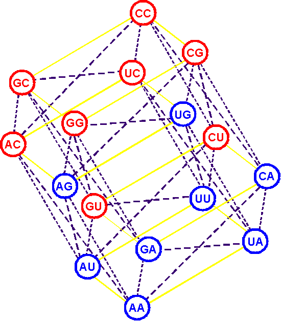
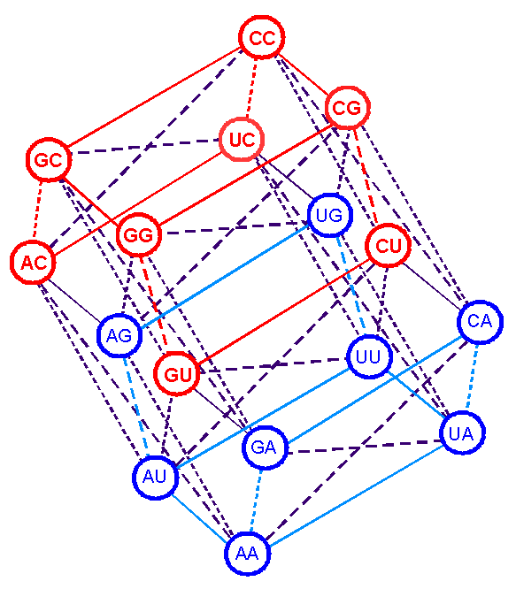
A. Single transitions |
||
|
Transitions C<-->G, U<-->A are united into
parallelograms lying in a horizontal plane (Fig. 4) |
Transitions C<-->U, G<-->A
form parallelograms located in a vertical plane. |
Transitions C<-->A, U<-->G
connect
diagonals of parallelograms of the previous two types |
|
|
|
|
|
Fig. 4. Single transitions
C<-->G, U<-->A on structure of the doublet code. |
Fig.
5. Single transitions
C<-->U, G<-->A
in the spatial structure of the doublet code. |
Fig.
6. Single transitions
C<-->A, U<-->G
in the spatial structure of the doublet code. |
|
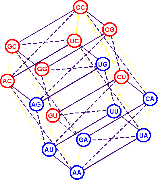
B. Rumer’s transformation |
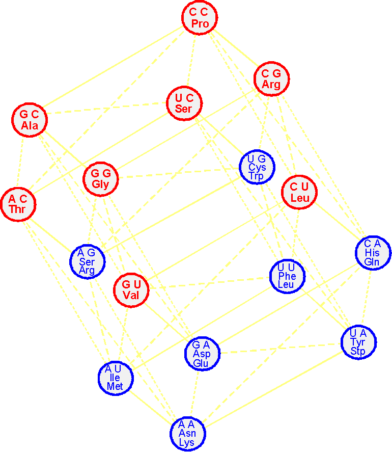
C. The
compact arrangement of amino acids. Cycles |
|||
|
The doublets, coding for one and two
amino acids and related by Rumer’s rule (C<-->A, G<-->U), occupy a symmetrical position
in the hypercube, for example: AC<-->СA, GC<-->UA, GG<-->UU, GU<-->UG, etc. |
|
|||
|
|
|
|||
|
Fig. 7. The position of doublets connected by
transformation of Rumer, in a doublet structure
of the genetic code. |
Fig. 8. The location of amino acids in the spatial structure of the
doublet genetic code. |
After becoming acquainted with the
principles of construction of the spatial structure of the doublet code it will be easy to understand how to construct the spatial structure of the triplet genetic code (section 2.3.).